Monday, June 12, 2017

Lab Report : Introduction to Light Microscopy

Hello everyone! :)

As I told you before, in this year we made nine different experiments in our biology lab classes. And I'll share the important parts of my lab reports regularly with you through this blog. As a result, you can see what we did and learned in this year in BIO 106. In addition, I think, these reports can give you some advice about how biological lab reports should be written, etc. I hope, they will be useful for you :) 

Here is the first experiment of this semester: 

INTRODUCTION TO LIGHT MICROSCOPY

THE AIM OF THE EXPERIMENT:

The aims of observations in this experiment were to specify different eukaryotic cell structures of animal and plant cells, to determine their morphological properties which can be observed under a light microscope, to estimate the sizes of animal and plant cells and, to determine and compare the structural and basic differences between Gram (+) and Gram (-) bacteria.

INTRODUCTION:

Until the invention of the first microscope in the seventeenth century, scientists were not be able to see and observe the cells and other tiny living and non-living things which couldn’t be seen with naked eyes. Light microscopes were the first microscopes which is used by scientists to see the complex structure that underlies all living things. After that, scientists developed different technologies such as Fluorescence Microscopes, Confocal Microscopes, Transmission and Scanning Electron Microscopy to observe specific structures of different cells.


The Magnification of a microscope is the measurement of enlarging of a sample’s image under a microscope and total magnification of a compound light microscope which includes two different lenses: objective lens and ocular lens, can be calculated by multiplying both two lenses’ magnification degrees. Resolution is described as the ability of a microscope to distinguish the details of the samples and the power of resolution (resolving power) of a microscope is determined by the numerical aperture of its objective. The difference in the intensity of light, between the image of a sample and its background relative to the intensity of the overall background is defined as the “Contrast” of a microscope and when a specimen is transparent or lack of color, the contrast of this specimen is needed to improve by using dyes, etc. In addition, the immersion oil is a synthetic oil which is used by scientists to increase the resolving power of a microscope and to arrange the brightness of the image of the specimen which is observed through the microscope.


Under a light microscope, some structural differences between animal, bacterial and plant cells can be observed. None of animal cells has a cell wall, however the plant cells and most of the bacterial cells have their own special type of cell walls. In addition, plant cells have an ordinary structure together and their cells’ shapes are mostly like a quadrilateral, however bacterial and animal cells doesn’t have a specific shape and they don’t build an ordinary view together. On the other hand, the chloroplast excited by the light can only be seen in plant cells.


By using Gram Staining Method, Gram(+) and Gram(-) bacteria can be distinguished with crystal violet dye. Gram(+) bacteria retain the dye and can be seen purple, Gram(-) bacteria cannot retain the dye inside because of its second outer membrane and appear pink.

METHODS:

Preparing and Cleaning The Microscope:

·         To be able to use 100X lens with immersion ail properly, the 100X lens was cleaned by using Isopropanol and a tissue paper at the beginning and at the end of the experiment.
·         The microscope was prepared according to the directions of the Lab Assistant and focused for both eyes of performers for each observation individually.

Observation of The Printed Letter “e”:

·         A printed black “e” letter was put on a microscope slide.
·         Two or three drops of water were dripped on the letter properly by using a Pasteur pipette.
·         A coverslip was placed on the letter and water on the microscope slide.
·         To prevent and reduce the bubble formation between the coverslip and the paper, the back side of a Pasteur pipette was pressed to the coverslip gently.
·         The microscope slide was placed on the microscope stage.
·         The letter “e” was centered and the microscope was focused for both eyes.
·         For observing the letter “e”, three different objective lenses were used. Firstly, 4X lens, and then 10X and 40X lenses were used.
·         For each step, the observations under different lenses were made and drawn to the lab notebook.
·         The pictures of the letter under different lenses were taken.

The Observation of Buccal Smear:

·         By using a Pasteur pipette, one or two water droplets were placed on a slide
·         A toothpick was used to get epithelial cell samples from the mouth of the performer’s partner (E.D) by scrapping the toothpick inside of his cheek.
·         The toothpick with cell samples was stirred into the water droplets on the slide.
·         A coverslip was placed on the sample.
·         One or two droplets of a dilute methylene blue solution were added to one edge of the coverslip on the sample.
·         The dye was drawn under the coverslip by using a tissue paper.
·         The dyed specimen was observed under three different objective lenses. Firstly, 10X lens was used. After that, 40X and 100X lenses were used.
·         When 100X lens was used, one or two droplets of immersion oil were dripped on the coverslip.
·         The observations under different lenses were made and drawn and their pictures were taken.

Using The Hemocytometer:

·         By using another toothpick, a new epithelial cell sample was gotten from the performer’s mouth (K.E.Ç).
·         One or two water droplets were placed on a glass slide with a hemocytometer.
·         The toothpick with new sample was stirred into the water droplets on the slide.
·         A coverslip was placed on the sample.
·         Under the coverslip, one droplet of methylene blue was added to the sample.
·         The glass slide was placed on the microscope stage.
·         The samples were observed under 10X and 40X magnifications.
·         By using the squares and lines on hemocytometer under 40X lens, the diameter of the field of view and the diameter of one cell were calculated.
·         The calculations and the observations were written and drawn.

The Observation of Elodea Cells:

·         The samples from Elodea leaves were taken from the Lab Assistant.
·         The Elodea sample was placed on a slide and one or two droplets of water were placed on the sample.
·         A coverslip was placed on the sample.
·         The sample was observed under 10X, 40X and 100X magnifications.
·         During the observations under 40X magnification, the horizontal and lateral sizes of the Elodea cells were calculated, according to the calculations in epithelial cell samples under 40X.
·         For the observation under 100X magnification, one droplet of immersion oil was placed on the coverslip.
·         The observations for each step and the calculations under 40X were written, drawn and their pictures were taken.

The Observations of Gram(+) and Gram(-) Bacteria:

·         Two different bacteria samples were taken from the Lab Assistant on prepared microscope slides.
·         The one labeled as “only E.coli” was observed under 4X, 10X, 40X and 100X magnifications.
·         For the observation under 100X, one droplet of immersion oil was placed on the coverslip of the sample.
·         The observations for each magnification were written and drawn.
·         The other slide with the label “E. coli + B.subtilis” was observed under 4X, 10X, 40X and 100X magnifications.
·         Immersion oil was used for the observation under 100X magnification.
·         All observations under each magnification were written and drawn.

DISCUSSION:

During the observations of this experiment, some specific differences between animal, bacterial and plant cells could be observed, as expected at the beginning of the experiment. In addition to that, estimating the sizes of animal and plant cells, determining the morphological properties of these cells and making a structural comparison between Gram(+) and Gram(-) bacteria were the other expectations of this experiment.

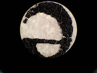
For these purposes, firstly a printed letter “e” was observed under 4X, 10X and 40X magnifications. As can be observed in Figure 1 and Figure 2, the images of the letter “e” were inverted and reversed. The causes of this altered view of the letter are the focal length of the objective lens and the lens’ curvature. The focal length of the objective lens of a microscope is very short and after the light passes through the printed “e”, the light also passes the objective lens of the microscope and the focal point of the objective lens. As a result of these steps, the images are inverted and reversed.

As expected at the beginning of the experiment, under different magnifications, the images were not in the same size. As long as the power of magnification of the lenses increased, the size of the images which were observed also increased. However, the directions of the movements of the images were the same for each power of lenses and they were to the opposite direction of the sample’s movement, because the objective lens of the microscope inverts the image of the sample.

As it can be observed, by using methylene blue to stain the buccal smear, the nucleus and the organelles of the epithelial cells which contains nucleic acids such as ribosomes and mitochondria could be observed. The methylene blue dye interacts and dyes the components of cells which contains nucleic acids darker than the other parts of the cell. The mitochondria have their own DNA and RNA molecules inside. In addition to that, ribosomes consist of rRNAs and they also contain nucleic acids to stain with methylene blue. On the other hand, on the rough Endoplasmic Reticulum in the cells there can be observed ribosomes, too and the Endoplasmic Reticulum is placed around the nucleus in the cells. As it can be seen in Figure 6, as a result of ER ribosomes’ existence, the density of the ribosomes around the nucleus are higher than other parts of the cell. If the buccal smear samples wouldn’t be dyed or stained with methylene blue, these observations couldn’t be made under a light microscope, because of the transparent existence of the epithelial cells. 


In the figure, it can be observed that because of their tetragonal shaped cell walls, plant cells have an ordinary structure in their tissues. In addition to that, there is no empty place between the cells of this sample and, the thickness of the cell walls are not the same for each cell in the sample, because of the difference between their lifetimes. On the other hand, there can be also seen the transportation channel which consists of Xylem and Phloem cells of the Elodea Leaf sample, in the Figure.

In the figure, the plant cells can be seen in more detail and the chloroplasts of the cells can also be seen during their movement because of the light excitation. However the nucleus of these plant cells couldn’t be seen under the 100X magnification.
The bacterial cells which were observed in the Gram Staining Experiment were in different colors. As it can be observed in the figure, the E.coli Bacteria have a line shaped structure and their color was pink. On the other hand, as it can be seen in Figure 14, the B.subtilis Bacteria have a circular cell structure and their color was violet. Because the Gram (+) B. subtilis Bacteria can retain the violet dye inside. However, the Gram(-) Bacteria E.coli cannot and appear in pink.


In the end of the experiment, it can be observed that animal cells, plant cells and bacterial cells have some differences in their structural basis such as having a cell wall for bacterial an plant cells, having an ordinary structure in their tissues for plant cells and having chloroplasts inside the their cells for the plant cells. In addition to that, the sizes of the cell samples were also different from each other. The buccal cell samples are bigger than the plant cells and the bacterial cells are the least. Because of that, the details of the observations of the inside cell structures were not the same for each cell type, too.

Friday, June 2, 2017

The beginning of Summer! :)

Hello everybody! :)

Finally, my first year at this department ended and it is the beginning of summer!! :)

I feel relief and completely happy, even though my GPA is not completely apparent. However, i hope that it will be enough to go Erasmus and in this situation, this is enough for me, too. To be honest, this year was not very easy for me. Because, I really didn't like some classes that I had to take and also the education system of some classes were not completely favorable to me. As a result, I couldn't stop asking questions about the system, about the classes and also about the department. And to be honest, I was thinking to change my department, for a while. However, I couldn't :D Because it is my DREAM JOB and it is not an easy decision to change it.

As a result, I really had some psychological problems at the end of the semester, because I wasn't happy. AND IT WAS A VERY BIG PROBLEM! I think, if I could only accept the situation that I had to be in it and could ask less questions about everything, I would be happier and probably more successful. However, I couldn't do that and because of that, I couldn't study enough for nothing but my German Literature Class :D I'm very very proud about my AA grade from this class and my Physics grade makes me also very happy :D But I can't say that for my Biology grade :D Even though, I haven't known my actual grade yet, I think that it won't be so nice. BUT DON'T BE AFRAID, I'M REALLY IN MOLECULAR BIOLOGY DEPARTMENT :D

I think, in this very first year, I learned a lot about the university, about the department and about how I should study for my classes etc. It was an adaptation year and I hope the next one will be better than this one :)

And now, LET THE SUMMER BEGIN! :) And here is the song for that : Summer!

SEE YOU SOON :)

LOVE YOU! :)

Saturday, April 29, 2017

About the Terrifying Month :D

Hello everybody! :)

It's me Back Again!:)

Approximately since the winter break, I couldn't find any time to write about the classes, lab days and other stuffs in my pretty hard education life :D

However, our second midterms and finals are coming like they are some parts of an invincible army! :D

In May, I have 11 exams ( 5 Midterms and 6 Finals), a lab report about the "Model Organisms" to give, a power point presentation for my Modern German Literature class and two different articles about Graph Theory Applications for Brain Cancer(glioma) to read for the Chapter I'll write.

You see, this month is completely terrifying and I really don't feel that I'm ready for it..

The exams start on this Tuesday and I REALLY have to study! I haven't written my lab report yet and haven't also prepared my power point presentation, too. And honestly, I have thousand pages to read about everything :D

I hope, I can handle it and start my summer holiday and my summer internship with having a good mood. Because, I really have to fix my GPA in this semester to be able to apply great universities for Erasmus next year! Actually, I'm kinda nervous about that, but I really want to handle it and show myself that I can do anything when I want to! But I don't know..
I hope that everything goes perfect! :)

After this terrifying month, I want to write everything about what I learned and made in my second semester! I'll share my lab reports, especially the introduction parts which take ALWAYS more than 4 hours to write! :D In addition, they really include great information about basic laboratory techniques which we learned in this semester! :)

So see you soon and alive (HOPEFULLY) :D

LOVE YOU <3

Kumsal

Sunday, January 22, 2017

Lab Diaries #Week1 :)

Hello everyone!

Yes, as you can understand from the title, our winter break has already begun and my lab days,too. :)
Honestly, it wasn't very easy to begin working in the lab because we couldn't meet with my lab professor to talk about my  position in the lab and about who will be the post-doc student that I'll work together, etc. because of the snowy weather!

Actually, I'm really in love with all the winter stuffs like snow, hot chocolate, sitting in front of the fireplace, etc. and I always feel better during these times. However, this time it was both: good and bad! Because, I was worried about my lab attendance!

To be honest, if I weren't in my first year at the university, I wouldn't be so worried about that, but it is obvious that I have no perfect skills to work at any lab(especially in a cancer/ gene regulation lab!) and I really don't know so much about my department,too. As a result, I always thought that what if the professor gives up from taking me to the lab..!

However, lucky me!, something like that didn't happened. In contrast, our professor was very interested about me and he talked about the future ( my future in the lab :D) very brightly! :) So, I'm very glad to say that : FINALLY, it has begun!

First thing I want to say about my first day in the lab is that I really didn't learn much during this semester! There are a lot of different things which I'm not familiar with and have to study for. Honestly, I even didn't know which machine using for what reason etc. Because of that, I took a lot of notes during the experiments and than I rewrite them to memorize what I've learned. After that, I decided to learn and read more about the machines and techniques which we are used during the lab and I'll share the articles that I read with you,too :)

In addition to that, my post-doc student (her name is Nalan and she is kind of a teacher for me too :) ) gave me some advice about what I should read and learn to be able to follow her experiments better. And during my first week I read one of the articles which she suggested to me. Here is the Link of the article : "The Hallmarks of Cancer". And I think that it was very useful to understand some basic structures of cancer! :)

In addition to read articles,of course I learned a lot during the experiments, too. Actually, the names of different enzymes, proteins and their effects etc. all the things that I saw, was completely new for me. Because of that I can make a huge list for what I've learned. However, as a summary maybe I can write some basic stuffs and procedures to see what I got from this experience, after my internship :

  • What is PCR and how can it be used? (I'll share a post about the technique, too :) )
  • Medium preparing for cell cultures
  • Oligo Annealing Protocol and its steps. (Oligo is a desalted custom synthesized DNA)
  • Different Buffers and how can they be prepared! (wash buffer, elution buffer, running buffer etc.)
  • "DOX" ----> for using over expression
  • Transformation - Step by step (with E.coli)
  • Transaction - Step by step (with Lenti Virus)
  • T4 Ligation
  • RNA Isolation - Step by Step
Of course, there can be some other things that I forgot to write but the basic things were these! :)

Next week, we will start some different experiments about her main project and I think those will be more interesting than these! :) I'm very excited! :)

So, see you next week! :)

LOVE YOU <3 :)


Kumsal

Sunday, December 25, 2016

Merry Christmas to you ALL :)


HELLO EVERYBODY!


I WISH YOU ALL A VERY MERRY CHRISTMAS!! :)

I HOPE YOU'LL HAVE GOOD TIME WITH YOU FAMILIES, FRIENDS etc.

I LOVE YOU ALL! <3 :)

SEE YOU SOON! :)

Kumsal

Saturday, December 24, 2016

Finals are COMING! :D :O


Hello everybody,

As you can see, my finals are coming and I'm very excited about them!(And a little bit terrified :D) 
Last week, I took my Physics and Maths finals and in this week, I'll take Chemistry, Chemistry Lab and Biology! 

Honestly, this semester I couldn't take what I expected before from Chemistry. Actually, the topics were interesting and at the beginning I was studying hard. However, after one month I lost my control over Chemistry and I didn't really catch up after that :D :/ To be honest, I know the topics and I studied for them before. But I have no practice and as a result, I cannot solve the problems during the exam! But, until Wednesday  I have 3 more days to study and I believe that I can figure out! :))

Actually, what I learned from this semester is that if I want to be a successful scientist and a successful student, I have to study regularly. Maybe not daily but weekly and for all subjects! Because it is obvious that I have to learn a lot of different things about different kind of subjects and this is the only way to learn all of them completely at the same time! :) As a result, I hope next semester I'll study and plan everything more carefully. :) To be honest, I HAVE TO :D

Because next semester I won't be free as much as this semester and it is possible that all my weekends will be completely full because of my debate tournaments! In addition to that, in January I'll begin to work in Lab and this will take time,too. And than, I want to study for Bridge,too. And of course I have to study for my lectures anyway :D So, if I cannot be planned as much as needed, it can be completely a disaster for me! :/ However, I cannot let that happen!! :)


So in the end, I have to go to study now! :D              

Take care of you! :)

And merry Christmas Eve to you ALL! :)

LOVE YOU! :) <3


Saturday, December 17, 2016

Last Month of 2016 🎄🎁

Hello everyone :)

We are at the end of a huge year and I'm really excited about the next one!

However, I believe that if you don't write a summary of a year, you cannot be prepared for the next one as much as you need! Because, it is obvious that people like to see and read what they could do and what they achieved in last year before they say "GOOD BYE!" to that and they can actually feel stronger when they see that they can do what ever they want if they want it ENOUGH!

And it works for me,too! As a result, here is my summary of 2016! :)

To be honest, this year was a productive year for me. I gained new skills and new hobbies, I got new friends and met a lot of perfect people in different parts of the year.

At the beginning of this year, I was in prep class and all I need to study was English! As a result, I could gain different skills during a semester. I had enough time to do something new! So, I could be an active member of our debate society! :) I could attend 6 or 7 tournaments and during these tournaments I met a lot of different people from different universities and got a lot of amazing friends! :)

In addition to that, I learned C and C++ Programming in this year. I joined in a class in Mimar Sinan Fine Arts University and I spent my free time to learn something new about computational biology! :) It was really entertaining and made a lot of fun! In addition, at the end of the semester, I made a project about protein interactions in different species and I learned so much about how should I prepare a project! It was perfect for me!

After that, I passed my proficiency test successfully! And this was very important for me, too ; because I really couldn't write or talk English more than some easy sentences! As a result, it is a success for me,too! :) And just before the test, we went our summer house in Bozcaada with my family for 4 days! :) It was a crazy and unplanned trip! However, it was completely amazing and relaxing!:)

After the second semester, my summer holiday was enough perfect for me,too. At the beginning, we organized a debate tournament in our Kilyos Campus (it is near the sea and perfect for a summer holiday!). It was really amazing and we spent great time together! :) After that, I went to Şile with my family to my uncles summer house! I was with my cousins and it was great,too.

In addition to that, I spent a lot of time with my best friends from my high school, because during the semester they weren't in Istanbul. One of them was in Germany, one of them was in Ankara and the other one was in Istanbul but we couldn't spend much time together because of our businesses! :D As a result, summer was a great opportunity for spending time together :)

At the beginning of this summer, we planned with my boyfriend to go on a vacation, However, we couldn't go :( Because his aunt and my brother got married in this summer! But we made some double dating stuffs with our friends, planned different things and could spend our summer holiday together perfect,too :D <3 :)

In addition to that, in summer I tried to join a Neurobiology Lab in Bahçeşehir University an for this purpose I made some very serious conversation first time in my life with different people. It was a successful meeting but the laboratory couldn't be opened, so I couldn't join to lab but; especially in this semester, I learned a lot from the professor in Bahçeşehir University. So, being able to meet him was a opportunity,too.

At the end of the summer, I made my very first class program in this year and it was completely excited! :D Even though, I didn't have any elective courses, it was something new :D

And than, this semester has begun!

To be honest, this semester was not perfect as I expected before, because the stuffs we learned were not very interesting as I wrote in another post and as a first year student, they were not easy,too!! As a result, I got some problems psychologically :( But I'm better now! :)

Besides the classes, I learned to play Bridge and joined the Bridge Club in this year and I found it really joyful! At the beginning, I just wanted to try how it works because my boyfriend loves this game! :) But now, I really love it,too. It's like chess with cards and makes me feel clever :D In addition, I believe it will be good for my CV and PhD application for universities in different countries in the future :)

As a success in this semester, I can say my lab attendance,too! In January, I'll begin to work in our "Cancer and Cell Regulation" Lab in our university and as a first year student, it is really an amazing chance for me!!! I'm very excited about that :)


So, this is my 2016! :) Of course I had bad things during the year,too.We all had them. But these are not important and talking about the good stuffs is always better! :)

I hope 2017 will be great as much as 2016 for all of us :)

Actually, writing all theses things makes me feel really better and you should try that,too! :) It is a good way to say good bye to 2016 :) LET'S TRY IT! :) MAKE YOUR OWN SUMMARY WITH PHOTOS :)

HAVE A NICE LAST MONTH :) <3

LOVE YOU! :) <3


Kumsal