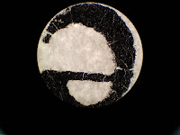
As I told you before, in this year we made nine different experiments in our biology lab classes. And I'll share the important parts of my lab reports regularly with you through this blog. As a result, you can see what we did and learned in this year in BIO 106. In addition, I think, these reports can give you some advice about how biological lab reports should be written, etc. I hope, they will be useful for you :)
Here is the first experiment of this semester:
INTRODUCTION TO LIGHT MICROSCOPY
THE AIM OF THE EXPERIMENT:
The aims of
observations in this experiment were to specify different eukaryotic cell
structures of animal and plant cells, to determine their morphological
properties which can be observed under a light microscope, to estimate the
sizes of animal and plant cells and, to determine and compare the structural
and basic differences between Gram (+) and Gram (-) bacteria.
INTRODUCTION:
Until
the invention of the first microscope in the seventeenth century, scientists
were not be able to see and observe the cells and other tiny living and
non-living things which couldn’t be seen with naked eyes. Light microscopes were the first
microscopes which is used by scientists to see the complex structure that
underlies all living things. After that, scientists developed different
technologies such as Fluorescence Microscopes, Confocal Microscopes,
Transmission and Scanning Electron Microscopy to observe specific structures of
different cells.
The
Magnification of a microscope is the measurement of enlarging of a sample’s
image under a microscope and total magnification of a compound light microscope
which includes two different lenses: objective lens and ocular lens, can be
calculated by multiplying both two lenses’ magnification degrees. Resolution is described as the ability of a microscope to distinguish the
details of the samples and the power of resolution (resolving power) of a
microscope is determined by the numerical aperture of its objective. The difference in the intensity of light, between the
image of a sample and its background relative to the intensity of the overall
background is defined as the “Contrast” of a microscope and when a specimen is
transparent or lack of color, the contrast of this specimen is needed to
improve by using dyes, etc. In addition, the immersion oil is a
synthetic oil which is used by scientists to increase the resolving power of a
microscope and to arrange the brightness of the image of the specimen which is
observed through the microscope.
Under
a light microscope, some structural differences between animal, bacterial and
plant cells can be observed. None of animal cells has a cell wall, however the
plant cells and most of the bacterial cells have their own special type of cell
walls. In addition, plant cells have an ordinary structure together and their
cells’ shapes are mostly like a quadrilateral, however bacterial and animal
cells doesn’t have a specific shape and they don’t build an ordinary view
together. On the other hand, the chloroplast excited by the light can only be seen
in plant cells.
By
using Gram Staining Method, Gram(+) and Gram(-) bacteria can be distinguished
with crystal violet dye. Gram(+) bacteria retain the dye and can be seen
purple, Gram(-) bacteria cannot retain the dye inside because of its second
outer membrane and appear pink.
METHODS:
Preparing and Cleaning The Microscope:
·
To be able to use 100X
lens with immersion ail properly, the 100X lens was cleaned by using
Isopropanol and a tissue paper at the beginning and at the end of the
experiment.
·
The microscope was
prepared according to the directions of the Lab Assistant and focused for both
eyes of performers for each observation individually.
Observation of The Printed Letter “e”:
·
A printed black “e”
letter was put on a microscope slide.
·
Two or three drops of
water were dripped on the letter properly by using a Pasteur pipette.
·
A coverslip was placed on
the letter and water on the microscope slide.
·
To prevent and reduce the
bubble formation between the coverslip and the paper, the back side of a
Pasteur pipette was pressed to the coverslip gently.
·
The microscope slide was
placed on the microscope stage.
·
The letter “e” was
centered and the microscope was focused for both eyes.
·
For observing the letter
“e”, three different objective lenses were used. Firstly, 4X lens, and then 10X
and 40X lenses were used.
·
For each step, the
observations under different lenses were made and drawn to the lab notebook.
·
The pictures of the
letter under different lenses were taken.
The Observation of Buccal Smear:
·
By using a Pasteur
pipette, one or two water droplets were placed on a slide
·
A toothpick was used to
get epithelial cell samples from the mouth of the performer’s partner (E.D) by
scrapping the toothpick inside of his cheek.
·
The toothpick with cell
samples was stirred into the water droplets on the slide.
·
A coverslip was placed on
the sample.
·
One or two droplets of a
dilute methylene blue solution were added to one edge of the coverslip on the
sample.
·
The dye was drawn under
the coverslip by using a tissue paper.
·
The dyed specimen was
observed under three different objective lenses. Firstly, 10X lens was used.
After that, 40X and 100X lenses were used.
·
When 100X lens was used,
one or two droplets of immersion oil were dripped on the coverslip.
·
The observations under
different lenses were made and drawn and their pictures were taken.
Using The Hemocytometer:
·
By using another
toothpick, a new epithelial cell sample was gotten from the performer’s mouth
(K.E.Ç).
·
One or two water droplets
were placed on a glass slide with a hemocytometer.
·
The toothpick with new
sample was stirred into the water droplets on the slide.
·
A coverslip was placed on
the sample.
·
Under the coverslip, one
droplet of methylene blue was added to the sample.
·
The glass slide was
placed on the microscope stage.
·
The samples were observed
under 10X and 40X magnifications.
·
By using the squares and
lines on hemocytometer under 40X lens, the diameter of the field of view and
the diameter of one cell were calculated.
·
The calculations and the
observations were written and drawn.
The Observation of Elodea Cells:
·
The samples from Elodea
leaves were taken from the Lab Assistant.
·
The Elodea sample was
placed on a slide and one or two droplets of water were placed on the sample.
·
A coverslip was placed on
the sample.
·
The sample was observed
under 10X, 40X and 100X magnifications.
·
During the observations
under 40X magnification, the horizontal and lateral sizes of the Elodea cells
were calculated, according to the calculations in epithelial cell samples under
40X.
·
For the observation under
100X magnification, one droplet of immersion oil was placed on the coverslip.
·
The observations for each
step and the calculations under 40X were written, drawn and their pictures were
taken.
The Observations of Gram(+) and Gram(-) Bacteria:
·
Two different bacteria
samples were taken from the Lab Assistant on prepared microscope slides.
·
The one labeled as “only
E.coli” was observed under 4X, 10X, 40X and 100X magnifications.
·
For the observation under
100X, one droplet of immersion oil was placed on the coverslip of the sample.
·
The observations for each
magnification were written and drawn.
·
The other slide with the
label “E. coli + B.subtilis” was observed under 4X, 10X, 40X and 100X
magnifications.
·
Immersion oil was used
for the observation under 100X magnification.
·
All observations under
each magnification were written and drawn.
DISCUSSION:
During the
observations of this experiment, some specific differences between animal,
bacterial and plant cells could be observed, as expected at the beginning of
the experiment. In addition to that, estimating the sizes of animal and plant
cells, determining the morphological properties of these cells and making a
structural comparison between Gram(+) and Gram(-) bacteria were the other
expectations of this experiment.
For these
purposes, firstly a printed letter “e” was observed under 4X, 10X and 40X
magnifications. As can be observed in Figure 1 and Figure 2, the images of the
letter “e” were inverted and reversed. The causes of this altered view of the
letter are the focal length of the objective lens and the lens’ curvature. The
focal length of the objective lens of a microscope is very short and after the
light passes through the printed “e”, the light also passes the objective lens
of the microscope and the focal point of the objective lens. As a result of
these steps, the images are inverted and reversed.
As expected at the
beginning of the experiment, under different magnifications, the images were
not in the same size. As long as the power of magnification of the lenses
increased, the size of the images which were observed also increased. However,
the directions of the movements of the images were the same for each power of
lenses and they were to the opposite direction of the sample’s movement,
because the objective lens of the microscope inverts the image of the sample.
As it can be
observed, by using methylene blue to stain the buccal smear, the
nucleus and the organelles of the epithelial cells which contains nucleic acids
such as ribosomes and mitochondria could be observed. The methylene blue dye interacts and dyes the components of cells which
contains nucleic acids darker than the other parts of the cell. The mitochondria have their own DNA and RNA molecules inside. In addition to
that, ribosomes consist of rRNAs and they also contain nucleic acids to stain
with methylene blue. On the other hand, on the rough Endoplasmic Reticulum in
the cells there can be observed ribosomes, too and the Endoplasmic Reticulum is
placed around the nucleus in the cells. As it can be seen in Figure 6, as a result of ER ribosomes’ existence, the
density of the ribosomes around the nucleus are higher than other parts of the
cell. If the buccal smear samples wouldn’t be dyed or stained with methylene
blue, these observations couldn’t be made under a light microscope, because of
the transparent existence of the epithelial cells.
 In the figure, it
can be observed that because of their tetragonal shaped cell walls, plant cells
have an ordinary structure in their tissues. In addition to that, there is no empty place between the cells of this sample
and, the thickness of the cell walls are not the same for each cell in the
sample, because of the difference between their lifetimes. On the other hand,
there can be also seen the transportation channel which consists of Xylem and
Phloem cells of the Elodea Leaf sample, in the Figure.
 In the figure, the
plant cells can be seen in more detail and the chloroplasts of the cells can
also be seen during their movement because of the light excitation. However the nucleus of these plant cells couldn’t be seen under the 100X
magnification.
The bacterial
cells which were observed in the Gram Staining Experiment were in different
colors. As it can be observed in the figure, the E.coli Bacteria have a line
shaped structure and their color was pink. On the other hand, as it can be seen
in Figure 14, the B.subtilis Bacteria have a circular cell structure and their
color was violet. Because the Gram (+) B. subtilis Bacteria can retain the
violet dye inside. However, the Gram(-) Bacteria E.coli cannot and appear in
pink.
In the end of the
experiment, it can be observed that animal cells, plant cells and bacterial
cells have some differences in their structural basis such as having a cell
wall for bacterial an plant cells, having an ordinary structure in their
tissues for plant cells and having chloroplasts inside the their cells for the
plant cells. In addition to that, the sizes of the cell samples were also
different from each other. The buccal cell samples are bigger than the plant
cells and the bacterial cells are the least. Because of that, the details of
the observations of the inside cell structures were not the same for each cell
type, too.


No comments:
Post a Comment