Friday, June 16, 2017

Lab Report: Cellular Fractionation

Hello everybody!

How are you? :)

In this week, I'm really busy in the lab and because of that, I couldn't write what we've done in the lab, yet. Yesterday, we made Nuclear Fractionation to some Bacteria to use their proteins  and today, to separate the proteins by using Western Blot Technique! It is a very long process and to follow truly, I read an article about the technique. At the weekend, maybe I can share a post about the Nuclear Fractionation Protocol that we used and an other post about Western Blotting :)

However, because I cannot write these posts before the whole process ends, today I will share my lab report about cellular fractionation by using homogenization technique. I think, it can be useful to read before reading the Nuclear Fractionation Protocol :)

Here it is :)

INTRODUCTION:

Centrifuge is a laboratory machine which is used for separating and isolating different substances from each other by using a motor which is able to spin the substances which are in liquid phase, at high speed. Differential Centrifugation is also one of the techniques for separating certain organelles from the cells by using their size and density differences.
Centrifuge machines have different types of rotors such as swinging bucket and fixed angle rotors. Swinging-bucket rotors can swing the sample tubes into a horizontal plane during the centrifugation process. On the contrary, fixed-angle rotors have a particular angle to fix the tubes during the centrifugation.To explain the properties of centrifuge machines, there are RPM and RCF values. RPM (Revolution per Minute) value shows the speed of the revolution that the rotor of the machine can reach and it is independent of the size of the rotor. However, RCF ( Relative Centrifugal Force) is the value of the force which is exerted on the samples in the rotor as a result of the revolution of the rotor and its value depends on the size of the rotor.


At the end of the centrifugation process, the substances in the tube separate from each other according to their sizes and densities. The smaller and less dense components move to the top of the tube and are called “supernatant”, and the larger and more dense components move to the bottom and are called “pellet”. During the cellular fractionation, densities and sizes of the needed components and environmental factors such as temperature and pressure have to be considered to obtain true and utilizable components from the sample.


Homogenization is a process in which the plasma membranes of the cells are ruptured and the cells are broken open. As a result, all the contents of the cells can be released. During this process, the cells are placed in an isotonic buffer and the resulting mixture is called “homogenate” which contains different, large and small components of the cells such as organelles, metabolites and enzymes. To homogenate cells and tissues, high frequency techniques(sonication), high pressure to make the cells move through a small hole(French press) and techniques to shear the cells(mechanical) can be used to break the cell membranes and soft detergents(detergent lysis) can be used to make holes on the cell membranes.

The centrifugation process and its results are affected by different properties such as RCF value, the duration of centrifugation, the shapes, sizes and densities of the cell samples, the density and the viscosity of the medium solution and, the column of suspension’s length. It is needed to be careful about the balance of the samples in the rotors and the lids of the tubes and the centrifuge have to be closed carefully and, until the centrifuge reaches the maximum speed, the samples shouldn’t be left.

AIM:
The aim of the experiment was to obtain mitochondria of the liver cells by using centrifugation technique.

METHODS:

1.      Homogenization: 
·         From an ice bucket, 1gr rat liver in a 50ml centrifuge tube was taken.
·         10ml of 0.25M sucrose solution was added in the tube.
·         By using homogenizer, a colloidal mixture of the rat liver and sucrose solution was prepared.
·         The homogenizer was used until there wasn’t any apparent liver piece in the tube.

2.      First Centrifugation:
·         A Table Top Centrifuge (swinging bucket) with RCF = 800g and RPM= 2037 values was used in the first centrifugation.
·         The centrifuge was arranged before the experiment to 4oC.
·         The 50ml falcon with 10ml mixture of liver and sucrose solution was placed in the rotor of centrifuge.
·         To balance the rotor during the process, the samples in the falcons were placed just opposite sides of each other.
·         The centrifuge was activated.
·         The machine was waited until it reached the maximum speed.
·         After 5 minutes, the sample was taken from the machine and it was observed that the pallet was at the bottom and the supernatant was at the top of the falcon.
·         The supernatant part of the sample was poured into a 15ml centrifuge tube to be used in the second centrifugation.
·         The pallet wasn’t used.

3.      Second Centrifugation:
·         To centrifuge at a higher speed, a High-Speed Centrifuge (fixed-angle) was used.
·         To determine the true RPM value that corresponds the needed RCF value, the instruction manual of the centrifuge was used.
·         To get the RCF = 5000g value, RPM = 5500 was calibrated on the machine.
·         To prepare a proper 15ml falcon with sucrose solution for balancing the sample in the rotor, the scales was used.
·         The centrifuge was arranged before the experiment to 4oC.
·         The adaptors with falcons were placed in the centrifuge and the lid was closed.
·         The centrifuge was activated.
·         The machine was waited until it reached the maximum speed.
·         After 15 minutes, the sample was taken from the machine and it was observed that the pallet was placed on the wall of the tube.
·         The supernatant was poured into a new 15ml falcon without taking the pellet to be used in the third centrifugation
·         The pallet wasn’t used.

4.      Third Centrifugation:
·         To centrifuge at a high speed, a High-Speed Centrifuge (fixed-angle) was used.
·         To determine the true RPM value that corresponds the needed RCF value, the instruction manual of the centrifuge was used.
·         To get the RCF = 24000g value, RPM = 12500 was calibrated on the machine.
·         To prepare a proper 15ml falcon with sucrose solution for balancing the sample in the rotor, the scales was used.
·         The centrifuge was arranged before the experiment to 4oC.
·         The adaptors with falcons were placed in the centrifuge and the lid was closed.
·         The centrifuge was activated.
·         The machine was waited until it reached the maximum speed.
·         After 15 minutes, the sample was taken from the machine and it was observed that the pallet was placed on the wall of the tube.
·         The supernatant was taken by using 5ml serological maxipipette without touching the pellet.
·         The supernatant wasn’t used.
·         Into the falcon with the pellet, 5ml of 0.25M sucrose solution was added.
·         The vertex was used to resuspend the mitochondrial pallet.
 Mitochondrial suspension was obtained.

DISCUSION:
The aim of this experiment was to obtain mitochondria of the liver cells for using them in different researches by using centrifugation technique. For this purpose, firstly the liver tissue was homogenized in 10ml of 0.25M sucrose solution. After that, three different centrifugation processes were performed and at the end of the experiment, the mitochondria of liver cells were obtained as the pellet.


For cellular fractionation, liver tissue was chosen because liver cells have more mitochondria than other tissues. Because of their work in the body, these cells require more energy than other cells and as a result, in their cytoplasm there are a lot of mitochondria to produce energy.

To perform the centrifugation processes truly, a proper solution for the sample was used. 0.25M sucrose solution was chosen to be used for this experiment, because it is an isotonic and dense solution. In addition to that, sucrose is a covalent molecule and it doesn’t dissolve ionically in a solution. On the other hand, sucrose molecules have a resolving effect for the other molecules in the sample and as a result they help to separate the compounds of sample, too.

All steps of this experiment were carried out in relatively lower temperatures because, the proteins and protein based structures in the liver cells and their components such as organelles and enzymes could be affected by high temperatures. As a result, these proteins and protein based structures could be denaturated and nonutilizable in the room temperature. Because of that, the liver tissue was kept in an ice bucket and the centrifuges were optimized at 4oC during the centrifugation processes.

On the other hand, to determine the true RPM value for the centrifuge, the instruction manual was used. The instruction manual shows the true RPM value for the needed RCF value and it was for RCF=24000, RPM = 12500. After the centrifugation, by using the formula to convert the RCF value to RPM value, the needed RPM value was calculated mathematically, too. The result was almost the same = 12552.

By using cellular fractionation, it is not possible to obtain a complete pure organelle solution. Because this method based on size and density differences and during the centrifugation process, the compounds of the cells could not be separated accurately, especially in this short time interval. Because of that, to obtain purer results the methods which can target the needed component directly can be used, such as antibodies and magnetic beads to separate the components.

I hope, you've had a perfect and productive week and you will have an amazing weekend at the end :)

See you soon..

LOVE YOU <3

Kumsal

Tuesday, June 13, 2017

Online Courses About Biological Sciences :)

Hello everyone!

How are you? :)
I really want to say that I'm completely full of energy but I'm not :D I'm really tired and it is a hot summer day. But thankfully, it is not sweltry.
I woke up at 8.00 am. and as I told before, my summer internship began yesterday. Because of that, I will spend my day in the lab, today :) So, I need coffee now :D

Also, yesterday was a productive day for me! :) I came to lab at 11 am. and began to study Japanese again :D I tried to learn Japanese before but I couldn't be so successful about that. However now, I have the whole summer and I think, it is the time to try again! :) I started with memorizing Hiragana and than I will try to learn Katakana and Kanji. I use this book to study and I have also a Duolingo account to study on the road :D  I hope, it will be better than first time!

In addition, I began to read some books about evolution and biodiversity because in next semester, we have a lesson about these topics. I want to be prepared, at least I want to have an idea about the subtopics. For this purpose, I use Biology: Concepts and Connections. I think, it's very clear and easily understandable.

And, now we can talk about our real topic! Another summer goal of mine is to attend some online courses about biological sciences and I made a list of some courses which can be taken in this summer until my 3. semester begins and I enrolled them. I hope some of them can be useful  for you,too. Here is my list:

1) Introductory Human Physiology 
(created by Duke University)

I takes 10 weeks and it begins July 03. To get information and see the syllabus

2) Music as Biology: What we like to hear and why
(created by Duke University)

It takes 6 weeks and it bagan yesterday! To get information

3) The Science of Gastronomy 
(created by The Hong Kong University of Science and Technology)

It takes 6 weeks and it will begin June 20. I think, it is really exiting! To get Information

4) Introduction to Genetics and Evolution
(created by Duke University)

It takes 11 weeks and it will start June 19. Information!

5) Introduction to Forensic Science
(created by Nanyang Technological University)

It takes 8 weeks and it will start June 26. It is very amazing, too. To get information
....

There are more of them and maybe I can write a post about all of the courses that can be useful for us. However, in one summer I can follow only a few of them. Because of that, I only wrote 5 courses.

I hope, they will be interesting for you, too.

See you soon :)

LOVE YOU! <3

Drosophila Melanogaster as a Model Organism :)

Hi everyone! :)

Today, I'm gonna share a post about Drosophila Melanogaster and three different experiments in which Drosophila was used as a model organism.

This text is also one of my lab reports for BIO 106 and to be honest, I wrote it at only one night. Because of that, there can be some grammatical problems :D However, I hope, you will enjoy that and learn new things from the text :)

DROSOPHILA MELANOGASTER


According to the evolution theory, it is thought that all living cells have a common ancestor cell and during the evolution of all living things, most of the basic properties of this ancestor cell have been conserved. That means, all species have mostly similar genetic properties and the differences between them are the results of small changes in their genome. As a result of that, especially in experimental biology researches, different species can be used as a test subject instead of each other. However, a certain number of organisms and cells which have different advantages about different experimental properties such as rapid reproducing, simplicity and transparency, are widely used as biological models in different experiments. In addition to that, because these chosen and determined species are used commonly by different scientists for different experiments, they can be examined deeply and scientific knowledge about the genetic properties of these species is very detailed. As a result of their different benefits, these species such as Escherichia coli and Saccharomyces cerevisiae are used as model organisms in biological experiments especially which are about human biology.

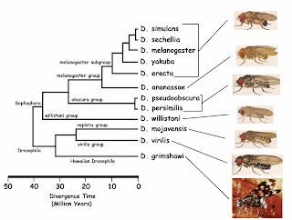
Drosophila Melanogaster is also one of these model organisms which are commonly used in experiments about human diseases and development stages. The usage of Drosophila in biological experiments have different advantages and disadvantages depending on the expectations from the experiments.

The most important benefit of Drosophila is that its genome was completely sequenced and published, and it contains a lot of conserved gene sequences which are similar with human genes. In addition to that, 75% of the genes which are related to different kinds of human diseases also can be find in its genome, too. As a result, Drosophila is commonly used as a model organism in drug design experiments for different human diseases.

The rapid life cycle of Drosophila is also an advantage of this model organism. Because from a single pair of this organism, it can be obtained more than a hundred of offspring which has identical genetic properties in about 10 days under laboratory conditions. In addition, to breed this organism under laboratory conditions is completely easier comparing to other species. They don’t need to have a complex diet to grow and they are not so expensive to study.


To have changing developmental stages such as embryo, larva, pupa and adult is a benefit of using Drosophila in different experiments. Because each stage of its development can be used for different researches such as about neuronal development and morphological changes. In addition to that, the adult stage of Drosophila has a complex systematic structure which has similar properties with human body systems and this property makes them also very useful for experiments about human development.

On the other side, Drosophila melanogaster has polytene chromosomal structure: its chromosomes are very big and they have light and dark colors of lines on them. As a result, its chromosomes can be observed under light microscope easily. And its body structures can be also observed with and without light microscope easily in different researches.

In addition to its advantages, Drosophila has some disadvantages for some biological experiments. One of its disadvantages is that even though they have similarities, the anatomy of Drosophila is actually very different. In addition to that, the fruit fly doesn’t have a complete adaptive immune system and they are not very useful for these type of experiments. And, Drosophila also doesn’t have some neurotransmitters and receptors which are important for neurological human diseases and this can be a disadvantage of using Drosophila instead of other species.


However, even though Drosophila Melanogaster has some disadvantages, its benefits are completely higher and it is commonly used in different kind of experiments about human diseases. One of these experiments, in which Drosophila was used is about the malignancy cancer investigations and different therapies for it. Even though some anatomical and physiological properties of Drosophila are quite different from humans, malignant tumors have a lot of similar effects on humans and Drosophila. The cells in both species firstly have changes in their developmental progress and their growth isn’t under control anymore. At the end, the tumor cells become immortal and cause the death of the patient. As a result of these similarities in stages of malignancy cancer in both species, different treatment options were tried on the Drosophila to be used in humans and by using these similarities and widely studied genomic properties of Drosophila, it was tried to understand the different molecular bases of malignancy.

Another experiment which was performed by using Drosophila Melanogaster is mapping the human cancer pathways. The human cancers develop through different stages such as mutations in cell – cycle, cell – death pathway and interactions about tumors, and to observe these steps of tumor formation easily, a model organism which has simple properties such as Drosophila was used. According to the results of some other experiments, by using the high ability in Drosophila to study interactions of tumor suppressors and oncogenes and, to generate tumor development and metastasis models; the hallmarks of cancer in Drosophila was determined, the pathways which promote the self – sufficiency was studied by using information about the effects of genes in Drosophila to self – sufficiency in growing and proliferation processes. And then, by using the different and well-studied cell signaling pathways of Drosophila, the effects of restraints against different processes such as cell proliferation, cell growth or the effects of blocking them was observed and the important cancer pathways such as RasACT and NothchACT were examined.


Drosophila Melanogaster is an appropriate model organism for modelling the neurodegenerative diseases and Parkinson’s disease is also one of the neurodegenerative diseases which were modelled by using Drosophila in an other experiment. The Parkinson’s disease is a disorder about movement and it is the result of losing the dopaminergic neurons in the substantia nigra part of the brain and also the result of filamentous intraneuronal inclusions formation. The gene α – synuclein is related to this disease and it is also effective on accumulation in Lewy bodies which are the filamentous intraneuronal inclusions. In order to express a genetic approach for Parkinson’s disease by using Drosophila, in this experiment firstly, the normal and mutant α – synuclein genes of Drosophila were expressed. After that, the loss of dopaminergic neurons in adulthood and Lewy bodies which contain α – synuclein were produced. In addition to that, a locomotor dysfunction was also generated in Drosophila and the results were observed and then used for modelling the disease by using the effects on Drosophila Melanogaster.

See you in another post! :)

LOVE YOU <3

Kumsal

Monday, June 12, 2017

Lab Report : Introduction to Light Microscopy

Hello everyone! :)

As I told you before, in this year we made nine different experiments in our biology lab classes. And I'll share the important parts of my lab reports regularly with you through this blog. As a result, you can see what we did and learned in this year in BIO 106. In addition, I think, these reports can give you some advice about how biological lab reports should be written, etc. I hope, they will be useful for you :) 

Here is the first experiment of this semester: 

INTRODUCTION TO LIGHT MICROSCOPY

THE AIM OF THE EXPERIMENT:

The aims of observations in this experiment were to specify different eukaryotic cell structures of animal and plant cells, to determine their morphological properties which can be observed under a light microscope, to estimate the sizes of animal and plant cells and, to determine and compare the structural and basic differences between Gram (+) and Gram (-) bacteria.

INTRODUCTION:

Until the invention of the first microscope in the seventeenth century, scientists were not be able to see and observe the cells and other tiny living and non-living things which couldn’t be seen with naked eyes. Light microscopes were the first microscopes which is used by scientists to see the complex structure that underlies all living things. After that, scientists developed different technologies such as Fluorescence Microscopes, Confocal Microscopes, Transmission and Scanning Electron Microscopy to observe specific structures of different cells.


The Magnification of a microscope is the measurement of enlarging of a sample’s image under a microscope and total magnification of a compound light microscope which includes two different lenses: objective lens and ocular lens, can be calculated by multiplying both two lenses’ magnification degrees. Resolution is described as the ability of a microscope to distinguish the details of the samples and the power of resolution (resolving power) of a microscope is determined by the numerical aperture of its objective. The difference in the intensity of light, between the image of a sample and its background relative to the intensity of the overall background is defined as the “Contrast” of a microscope and when a specimen is transparent or lack of color, the contrast of this specimen is needed to improve by using dyes, etc. In addition, the immersion oil is a synthetic oil which is used by scientists to increase the resolving power of a microscope and to arrange the brightness of the image of the specimen which is observed through the microscope.


Under a light microscope, some structural differences between animal, bacterial and plant cells can be observed. None of animal cells has a cell wall, however the plant cells and most of the bacterial cells have their own special type of cell walls. In addition, plant cells have an ordinary structure together and their cells’ shapes are mostly like a quadrilateral, however bacterial and animal cells doesn’t have a specific shape and they don’t build an ordinary view together. On the other hand, the chloroplast excited by the light can only be seen in plant cells.


By using Gram Staining Method, Gram(+) and Gram(-) bacteria can be distinguished with crystal violet dye. Gram(+) bacteria retain the dye and can be seen purple, Gram(-) bacteria cannot retain the dye inside because of its second outer membrane and appear pink.

METHODS:

Preparing and Cleaning The Microscope:

·         To be able to use 100X lens with immersion ail properly, the 100X lens was cleaned by using Isopropanol and a tissue paper at the beginning and at the end of the experiment.
·         The microscope was prepared according to the directions of the Lab Assistant and focused for both eyes of performers for each observation individually.

Observation of The Printed Letter “e”:

·         A printed black “e” letter was put on a microscope slide.
·         Two or three drops of water were dripped on the letter properly by using a Pasteur pipette.
·         A coverslip was placed on the letter and water on the microscope slide.
·         To prevent and reduce the bubble formation between the coverslip and the paper, the back side of a Pasteur pipette was pressed to the coverslip gently.
·         The microscope slide was placed on the microscope stage.
·         The letter “e” was centered and the microscope was focused for both eyes.
·         For observing the letter “e”, three different objective lenses were used. Firstly, 4X lens, and then 10X and 40X lenses were used.
·         For each step, the observations under different lenses were made and drawn to the lab notebook.
·         The pictures of the letter under different lenses were taken.

The Observation of Buccal Smear:

·         By using a Pasteur pipette, one or two water droplets were placed on a slide
·         A toothpick was used to get epithelial cell samples from the mouth of the performer’s partner (E.D) by scrapping the toothpick inside of his cheek.
·         The toothpick with cell samples was stirred into the water droplets on the slide.
·         A coverslip was placed on the sample.
·         One or two droplets of a dilute methylene blue solution were added to one edge of the coverslip on the sample.
·         The dye was drawn under the coverslip by using a tissue paper.
·         The dyed specimen was observed under three different objective lenses. Firstly, 10X lens was used. After that, 40X and 100X lenses were used.
·         When 100X lens was used, one or two droplets of immersion oil were dripped on the coverslip.
·         The observations under different lenses were made and drawn and their pictures were taken.

Using The Hemocytometer:

·         By using another toothpick, a new epithelial cell sample was gotten from the performer’s mouth (K.E.Ç).
·         One or two water droplets were placed on a glass slide with a hemocytometer.
·         The toothpick with new sample was stirred into the water droplets on the slide.
·         A coverslip was placed on the sample.
·         Under the coverslip, one droplet of methylene blue was added to the sample.
·         The glass slide was placed on the microscope stage.
·         The samples were observed under 10X and 40X magnifications.
·         By using the squares and lines on hemocytometer under 40X lens, the diameter of the field of view and the diameter of one cell were calculated.
·         The calculations and the observations were written and drawn.

The Observation of Elodea Cells:

·         The samples from Elodea leaves were taken from the Lab Assistant.
·         The Elodea sample was placed on a slide and one or two droplets of water were placed on the sample.
·         A coverslip was placed on the sample.
·         The sample was observed under 10X, 40X and 100X magnifications.
·         During the observations under 40X magnification, the horizontal and lateral sizes of the Elodea cells were calculated, according to the calculations in epithelial cell samples under 40X.
·         For the observation under 100X magnification, one droplet of immersion oil was placed on the coverslip.
·         The observations for each step and the calculations under 40X were written, drawn and their pictures were taken.

The Observations of Gram(+) and Gram(-) Bacteria:

·         Two different bacteria samples were taken from the Lab Assistant on prepared microscope slides.
·         The one labeled as “only E.coli” was observed under 4X, 10X, 40X and 100X magnifications.
·         For the observation under 100X, one droplet of immersion oil was placed on the coverslip of the sample.
·         The observations for each magnification were written and drawn.
·         The other slide with the label “E. coli + B.subtilis” was observed under 4X, 10X, 40X and 100X magnifications.
·         Immersion oil was used for the observation under 100X magnification.
·         All observations under each magnification were written and drawn.

DISCUSSION:

During the observations of this experiment, some specific differences between animal, bacterial and plant cells could be observed, as expected at the beginning of the experiment. In addition to that, estimating the sizes of animal and plant cells, determining the morphological properties of these cells and making a structural comparison between Gram(+) and Gram(-) bacteria were the other expectations of this experiment.

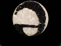
For these purposes, firstly a printed letter “e” was observed under 4X, 10X and 40X magnifications. As can be observed in Figure 1 and Figure 2, the images of the letter “e” were inverted and reversed. The causes of this altered view of the letter are the focal length of the objective lens and the lens’ curvature. The focal length of the objective lens of a microscope is very short and after the light passes through the printed “e”, the light also passes the objective lens of the microscope and the focal point of the objective lens. As a result of these steps, the images are inverted and reversed.

As expected at the beginning of the experiment, under different magnifications, the images were not in the same size. As long as the power of magnification of the lenses increased, the size of the images which were observed also increased. However, the directions of the movements of the images were the same for each power of lenses and they were to the opposite direction of the sample’s movement, because the objective lens of the microscope inverts the image of the sample.

As it can be observed, by using methylene blue to stain the buccal smear, the nucleus and the organelles of the epithelial cells which contains nucleic acids such as ribosomes and mitochondria could be observed. The methylene blue dye interacts and dyes the components of cells which contains nucleic acids darker than the other parts of the cell. The mitochondria have their own DNA and RNA molecules inside. In addition to that, ribosomes consist of rRNAs and they also contain nucleic acids to stain with methylene blue. On the other hand, on the rough Endoplasmic Reticulum in the cells there can be observed ribosomes, too and the Endoplasmic Reticulum is placed around the nucleus in the cells. As it can be seen in Figure 6, as a result of ER ribosomes’ existence, the density of the ribosomes around the nucleus are higher than other parts of the cell. If the buccal smear samples wouldn’t be dyed or stained with methylene blue, these observations couldn’t be made under a light microscope, because of the transparent existence of the epithelial cells. 


In the figure, it can be observed that because of their tetragonal shaped cell walls, plant cells have an ordinary structure in their tissues. In addition to that, there is no empty place between the cells of this sample and, the thickness of the cell walls are not the same for each cell in the sample, because of the difference between their lifetimes. On the other hand, there can be also seen the transportation channel which consists of Xylem and Phloem cells of the Elodea Leaf sample, in the Figure.

In the figure, the plant cells can be seen in more detail and the chloroplasts of the cells can also be seen during their movement because of the light excitation. However the nucleus of these plant cells couldn’t be seen under the 100X magnification.
The bacterial cells which were observed in the Gram Staining Experiment were in different colors. As it can be observed in the figure, the E.coli Bacteria have a line shaped structure and their color was pink. On the other hand, as it can be seen in Figure 14, the B.subtilis Bacteria have a circular cell structure and their color was violet. Because the Gram (+) B. subtilis Bacteria can retain the violet dye inside. However, the Gram(-) Bacteria E.coli cannot and appear in pink.


In the end of the experiment, it can be observed that animal cells, plant cells and bacterial cells have some differences in their structural basis such as having a cell wall for bacterial an plant cells, having an ordinary structure in their tissues for plant cells and having chloroplasts inside the their cells for the plant cells. In addition to that, the sizes of the cell samples were also different from each other. The buccal cell samples are bigger than the plant cells and the bacterial cells are the least. Because of that, the details of the observations of the inside cell structures were not the same for each cell type, too.